Guided implant planning · Muscat, Oman
Swiss-backed digital implant planning

The Future of Guided Surgery

Upload a CBCT. Receive a consultant’s report and a print-ready surgical guide — in as little as 3 days. Built for dentists across Iraq and the Gulf.

CR 1508999 Health Licence 1508999 Swiss & Italian systems Reply within 24h
A measured plate of four surgical guides made by Future Guide's own lab at one common scale, labelled with the dimensions measured from their files

Four guides printed in our own lab, shown at one scale. Every dimension is measured from the file the guide was printed from, none typed.

Six ways to start

Before you send anything

Send your CBCT, get a full plan

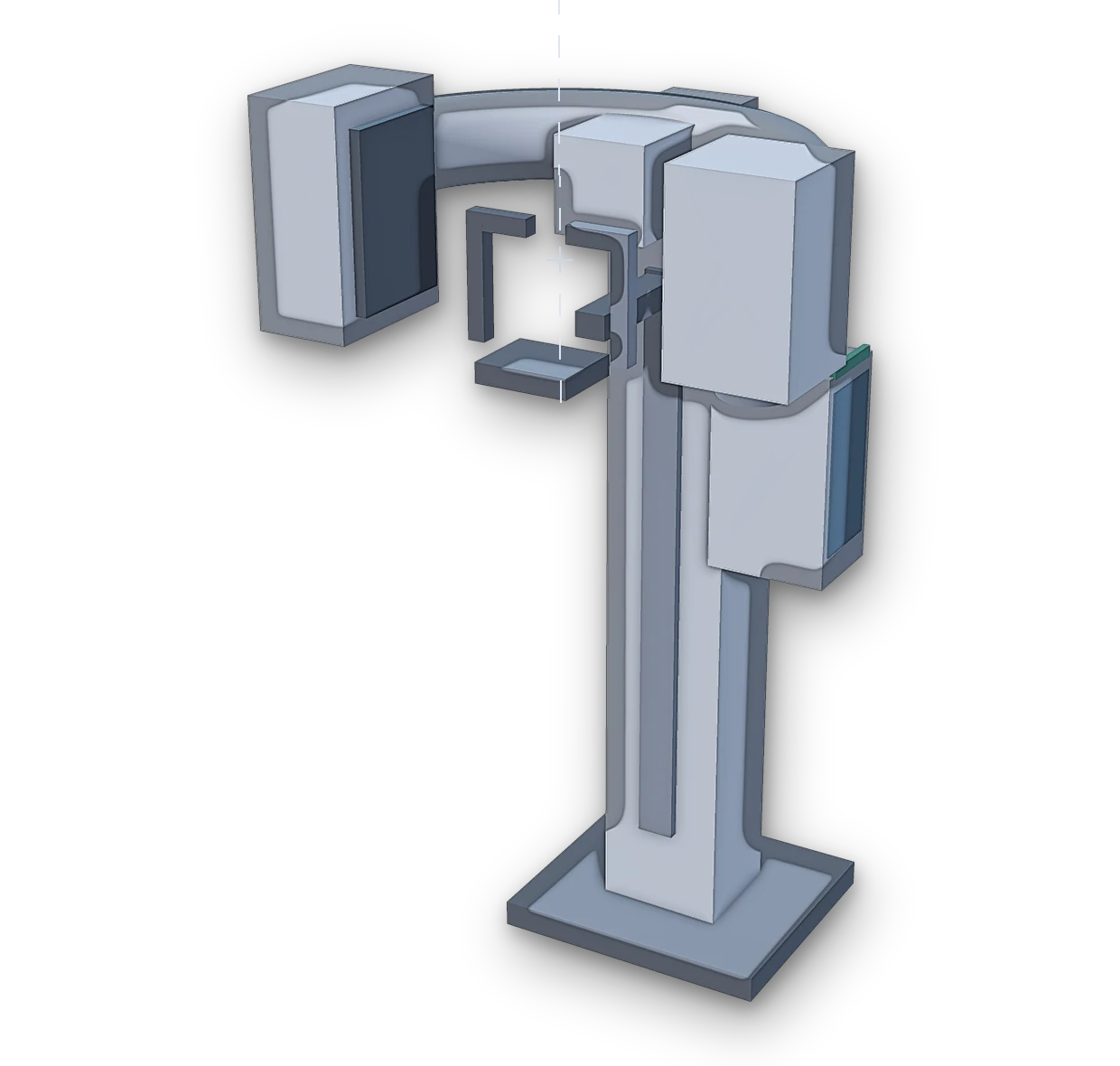
Free · nothing leaves your computer Drop in the scan and the impression — the reading comes back in seconds, in your own browser. Submit a case Check the file first — free Your clinic’s own CBCT, any make — we read only the file you send. Drawn, not photographed: no maker’s machine, no patient.
Get started

Send your first case today

Five steps, one upload. We confirm receipt the same working day.

Published cases, by other clinicians

Four published cases, planned the way we plan

Worked examples of the workflow this service is built on — the same sequence of guides, planned in the same software. They are here because they are instructive, not because they are ours.

Case one · whose case this is

Dr. Francescantonio Polito

Published in the clinical literature. Future Guide neither planned nor performed this case, and claims no part of it.

Full-Arch Immediate-Load Rehabilitation with a Sequential Guided Workflow

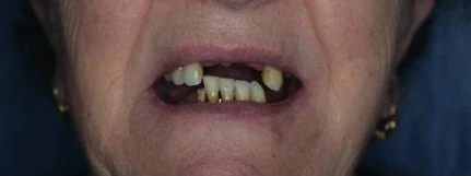
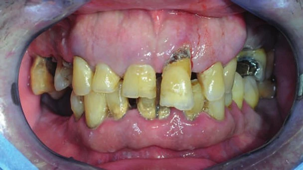
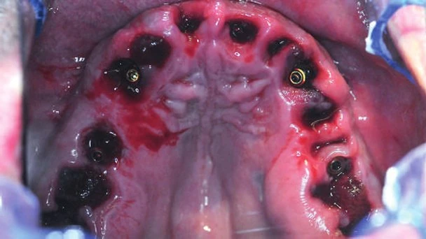
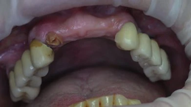
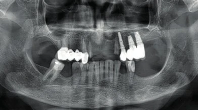
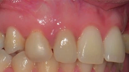
A 69-year-old woman in good general health presented with a severely compromised upper arch. Her clinical condition included multiple previous extractions, a residual denture damaged by extensive caries, several apical and periapical inflammatory processes, severe mobility of the remaining teeth, and a large area of palatal irritation caused by the removable prosthesis. Because of the resulting aesthetic and functional impairment, an implant-supported fixed rehabilitation was planned, with the upper arch treated as the first surgical phase.

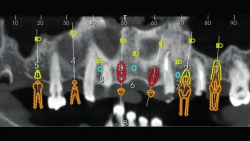
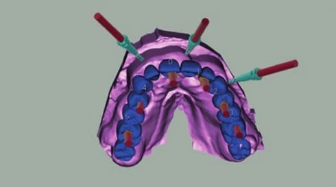
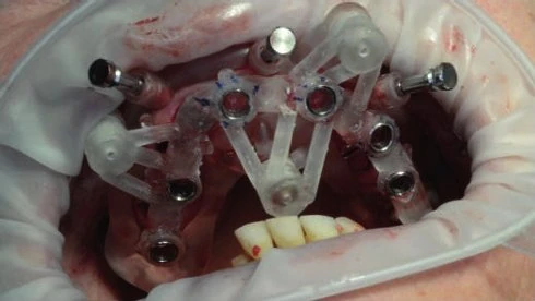
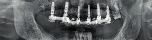
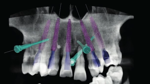
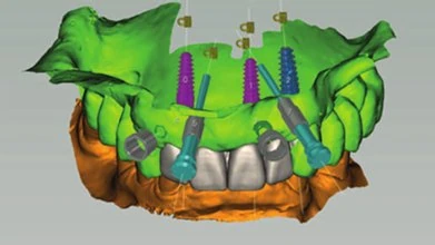
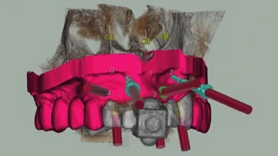
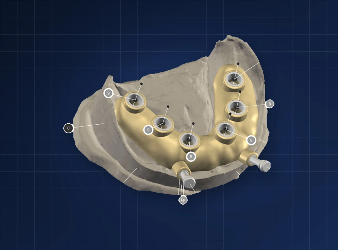
The digital workflow began by aligning the DICOM data with the STL files obtained from the intraoral scan. A diagnostic wax-up was imported into the guided-surgery software so that six implants could be planned according to the intended prosthetic result. The implant positions, dimensions, and prosthetic components were selected before surgery, and a preconstructed provisional restoration was incorporated into the treatment plan.

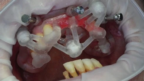
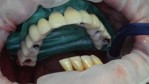
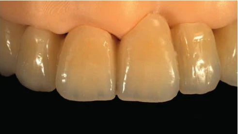
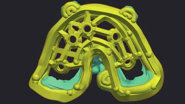
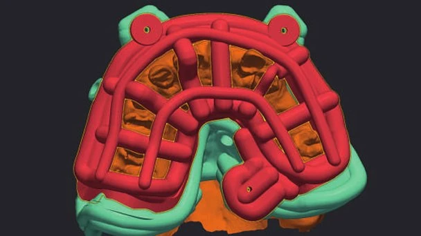
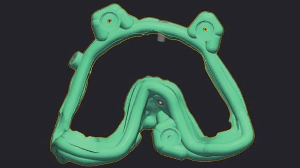
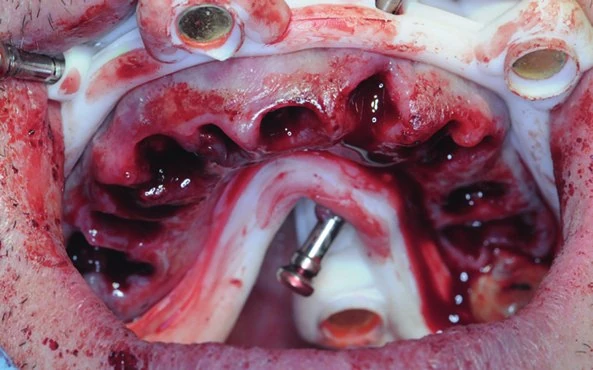
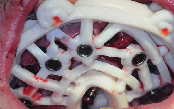
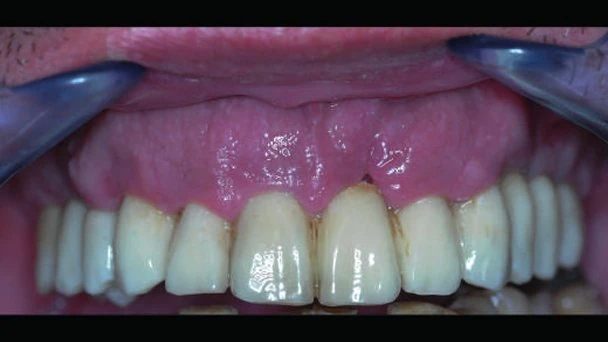
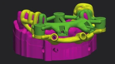
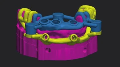
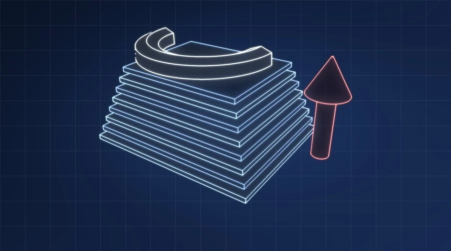
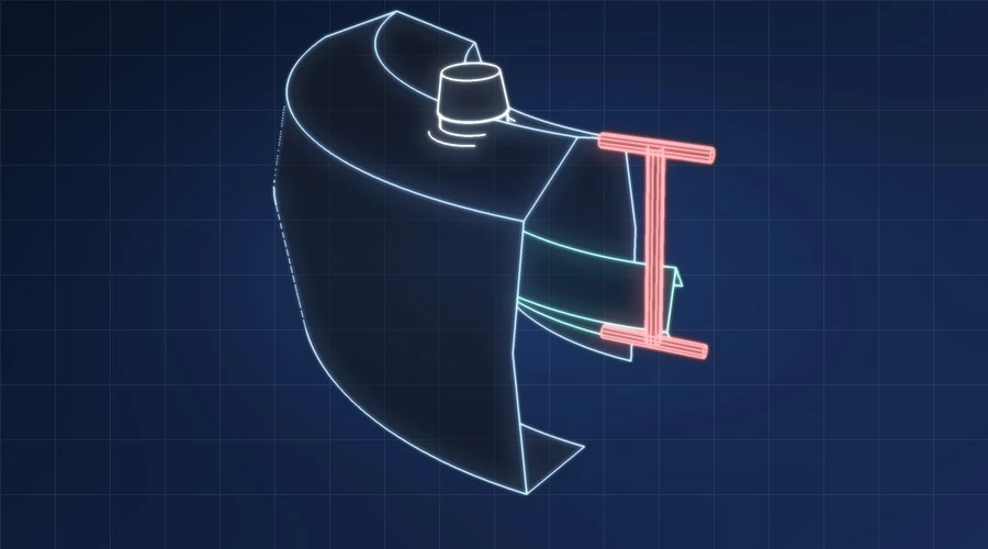
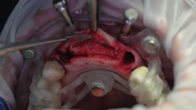
Using the SCULPT module, a sequence of predetermined templates was created for the extractions, limited osteoplasty, guided osteotomy preparation, implant insertion, and provisional restoration. A tooth-supported positioning guide established the location of an external base, which was stabilized with fixation pins and remained in place throughout the procedure. Magnetically coupled components were then exchanged in sequence while preserving the planned position. After guided implant placement and positioning of the prosthetic components, the prefabricated provisional restoration was connected at the end of the intervention.

This case demonstrates a complete digital and prosthetically driven workflow for full-arch immediate-load rehabilitation, from virtual planning and sequential guides to implant placement and delivery of the provisional restoration.

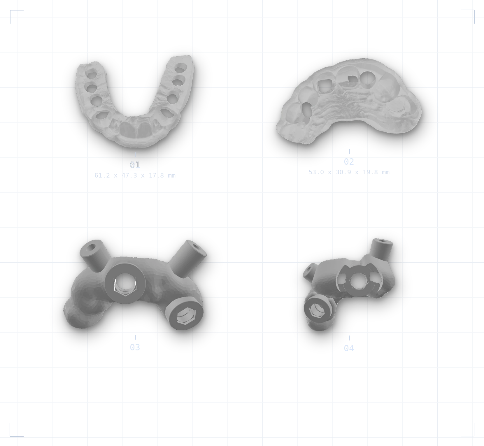
Figure 01 from the published case
Fig. 01
Figure 02 from the published case
Fig. 02
Figure 05 from the published case
Fig. 05
Figure 06 from the published case
Fig. 06
Figure 08 from the published case
Fig. 08
Figure 11 from the published case
Fig. 11
Figure 14 from the published case
Fig. 14
Figure 18 from the published case
Fig. 18
Figure 19 from the published case
Fig. 19
Case two · whose case this is

Dr. Michael Cerri

Published in the clinical literature. Future Guide neither planned nor performed this case, and claims no part of it.

Stackable Predetermined Guides for Immediate Post-Extraction Rehabilitation

This case presents the immediate-load rehabilitation of an entire post-extraction arch through a fully digital workflow. The clinical objective was to transfer the virtual plan accurately through the extraction, implant-placement, and provisional-restoration stages while maintaining a stable reference throughout treatment.

The predetermined-template concept used a bone-anchored base to reduce positional inaccuracy during implant insertion. Implant 3D Guide Design and the SCULPT module were used to create the separate portions of the guide system and to divide the temporary prosthesis into an aesthetic component and a supporting substructure. The resulting components could be used sequentially while maintaining the same planned reference position.

The patient received a screw-retained prosthesis on the same day. The clinical sessions lasted two hours in the morning and forty-five minutes in the afternoon. The case illustrates how preoperative digital design can support a predictable stackable workflow and shift a substantial part of the work from the surgical appointment to the planning stage.

Figure 05 from the published case
Fig. 05
Figure 09 from the published case
Fig. 09
Figure 12 from the published case
Fig. 12
Figure 13 from the published case
Fig. 13
Figure 15 from the published case
Fig. 15
Figure 18 from the published case
Fig. 18
Figure 19 from the published case
Fig. 19
Figure 23 from the published case
Fig. 23
Figure 25 from the published case
Fig. 25
Case three · whose case this is

Dr. Francescantonio Polito

Published in the clinical literature. Future Guide neither planned nor performed this case, and claims no part of it.

Selective Guided Crestal Osteoplasty with Immediate Implant Rehabilitation

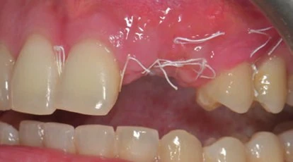
A 68-year-old woman required rehabilitation of the upper anterior region after losing a previously cemented prosthesis. The remaining root in position 12 and tooth 23 were severely compromised. The treatment plan included their extraction, post-extraction implants in positions 12 and 23, a third implant in position 21, and an immediately loaded provisional restoration.

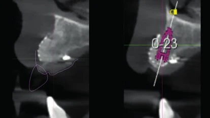
The initial digital workflow used Implant 3D and the SCULPT module. Because extensive metal-ceramic restorations complicated the alignment of the CBCT and model scan, a custom radiological device was designed to provide reliable reference points. A diagnostic wax-up then established the prosthetically correct implant positions. During planning, the team identified bone peaks that required corrective osteoplasty to create a more suitable ridge form in this highly aesthetic area.

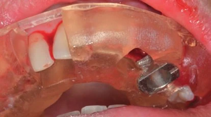
A dedicated bone-cutting guide was designed to make the reduction selective and controlled. Two virtual horizontal blade implants were positioned across the bone peaks to define the cutting paths and the corresponding piezoelectric bushings. A sequence of predetermined templates was then produced. A tooth-supported positioner established the external base, which was fixed with stabilization pins and remained in position throughout the procedure.

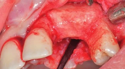
After the extractions and limited flap elevation, the bone-remodelling component guided the piezoelectric cutting action only over the planned peaks. A drilling template was then placed on the same stable base to guide osteotomy preparation and implant insertion. The prefabricated provisional restoration was connected in the planned position. At the later review, the book reports satisfactory hard- and soft-tissue integration and favourable tissue conditioning around the provisional restoration.
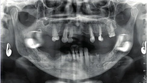
Figure 01 from the published case
Fig. 01
Figure 02 from the published case
Fig. 02
Figure 06 from the published case
Fig. 06
Figure 08 from the published case
Fig. 08
Figure 12 from the published case
Fig. 12
Figure 14 from the published case
Fig. 14
Figure 25 from the published case
Fig. 25
Figure 26 from the published case
Fig. 26
Figure 27 from the published case
Fig. 27
Figure 30 from the published case
Fig. 30
Figure 41 from the published case
Fig. 41
Figure 42 from the published case
Fig. 42
Case four · whose case this is

Dr. Nicola De Rosa

Published in the clinical literature. Future Guide neither planned nor performed this case, and claims no part of it.

Digitally Supported GBR Followed by Static Guided Implant Placement

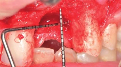
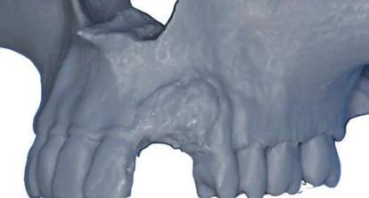
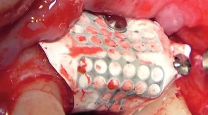
A healthy 22-year-old non-smoking patient presented with a marked tissue deficit in the 22-23 region. Surgical exposure revealed a bone deficiency of approximately 10 mm horizontally and 5 mm vertically. The treatment plan involved guided bone regeneration with a titanium-reinforced PTFE membrane, followed by implant placement and prosthetic rehabilitation.
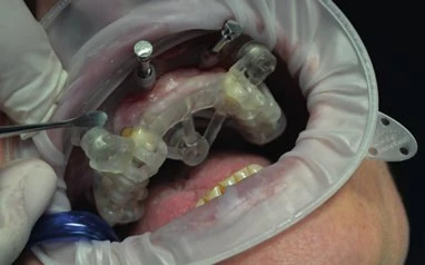
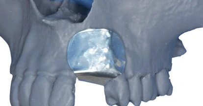
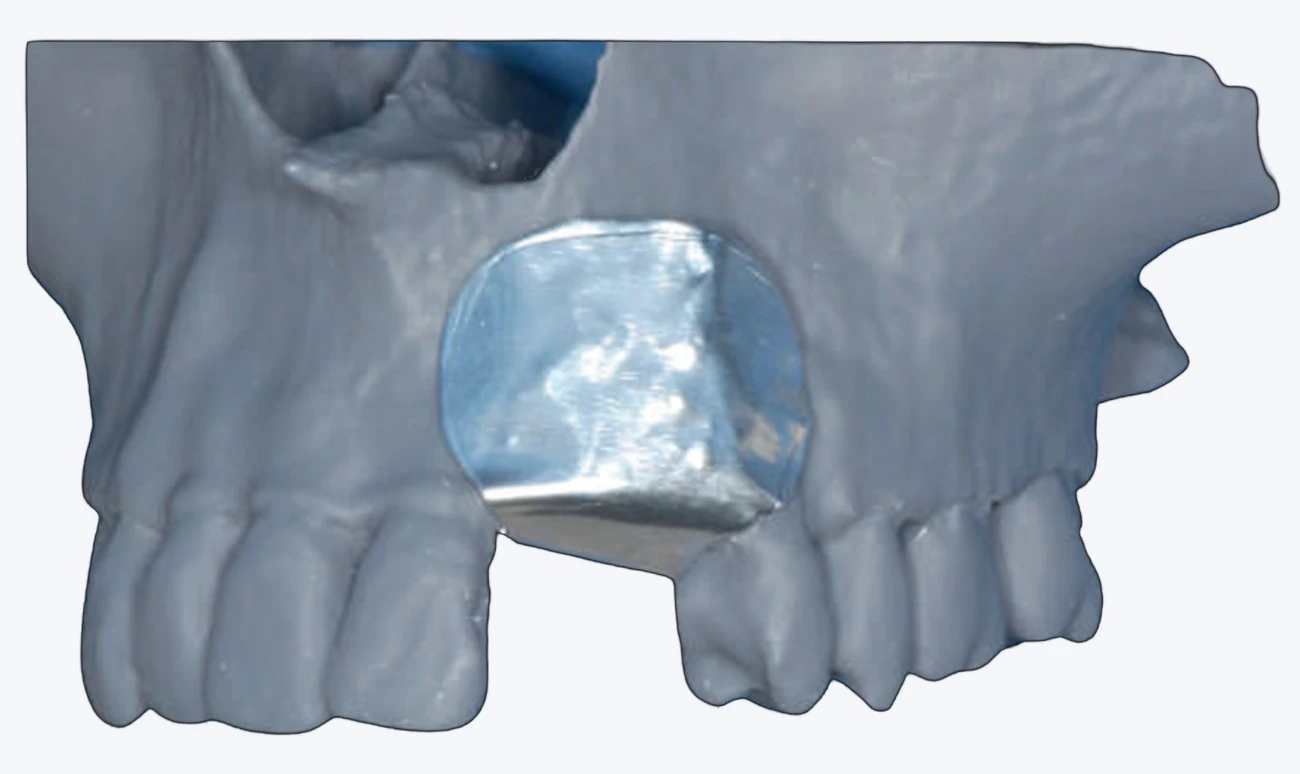
Before the regenerative procedure, a stereolithographic model of the maxilla was produced from the CBCT data. An aluminium template was created on this model and used intraoperatively as a support for shaping the membrane, reducing chairside working time. The graft consisted of 70% autogenous bone harvested from the ramus and 30% animal-derived bone. The book reports tension-free closure and favourable early healing.

After five months of GBR healing, a new CBCT and an intraoral scan were obtained. Implant position 23 was planned in Implant 3D according to a virtual diagnostic wax-up, and a tooth-supported static surgical guide was designed. On reopening, the regenerated bone was described as being of good quality. Guided osteotomy preparation and implant insertion were completed, followed by soft-tissue management. A fixed immediate-load restoration for positions 22 and 23 was placed five hours after surgery.

Figure 01 from the published case
Fig. 01
Figure 04 from the published case
Fig. 04
Figure 05 from the published case
Fig. 05
Figure 07 from the published case
Fig. 07
Figure 10 from the published case
Fig. 10
Figure 11 from the published case
Fig. 11
Figure 14 from the published case
Fig. 14
Figure 16 from the published case
Fig. 16
Figure 17 from the published case
Fig. 17
Figure 21 from the published case
Fig. 21
Figure 24 from the published case
Fig. 24
Figure 25 from the published case
Fig. 25

Your case, planned the same way

Send the CBCT and the scan and we will tell you what the plan has to solve before anything is drilled.

Services

Guided surgery services

Two services, one workflow. You send imaging; a consultant reviews it and we return either a written plan or a plan and the guide that executes it.

01

Consultative Report

A radiographic assessment of your case, written by a clinician.

What you upload

A CBCT scan of the region of interest, plus your case notes. Export the study as DICOM and upload the whole folder compressed into a single ZIP — not a screenshot and not a PDF report.

What to upload ZIP (DICOM) STL PLY OBJ max 1 GB

What you receive

  • Radiographic assessment of the region
  • Bone volume and density evaluation
  • Recommended implant position and angulation
  • Notes on anatomical structures to avoid
Delivery3 working days
Rush option24 hours
Free revisions1
02

Report + Surgical Guide

The full plan, and the physical guide file that carries it into the mouth.

What you upload

A CBCT — exported as DICOM and zipped as one folder — and an intraoral scan of the same arch as STL, PLY or OBJ, plus your case notes. Both are required: a guide cannot be designed from the CBCT alone.

What to upload ZIP (DICOM) STL PLY OBJ max 1 GB

What you receive

  • Everything included in the Consultative Report
  • A print-ready surgical guide file
  • Sleeve specification and drilling sequence
  • A digital preview for your approval before finalisation
Delivery5 working days
Rush option24 hours
Free revisions1
Clinical scope

Cases we plan

Beyond single-unit planning, we take on the cases that need the most control.

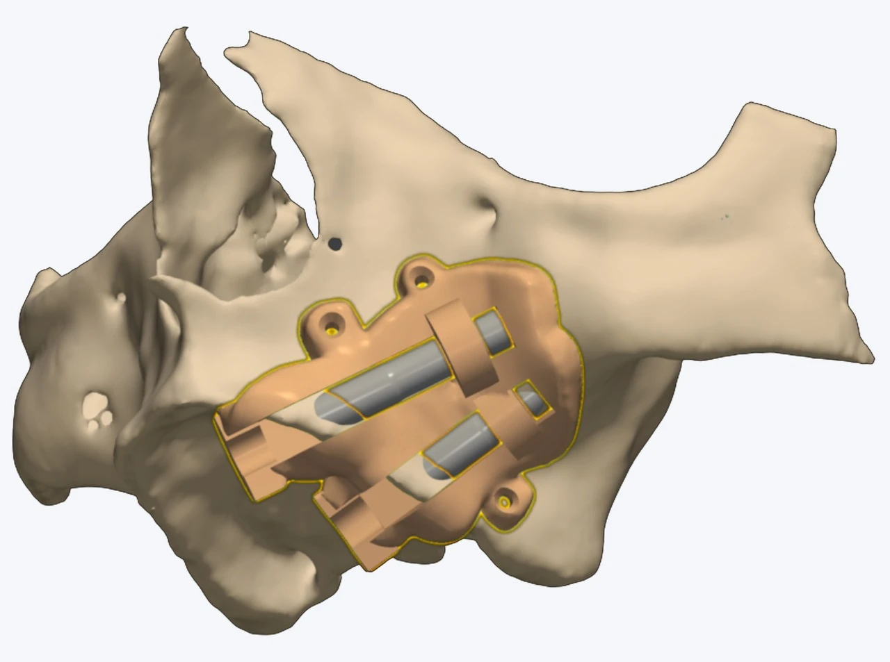
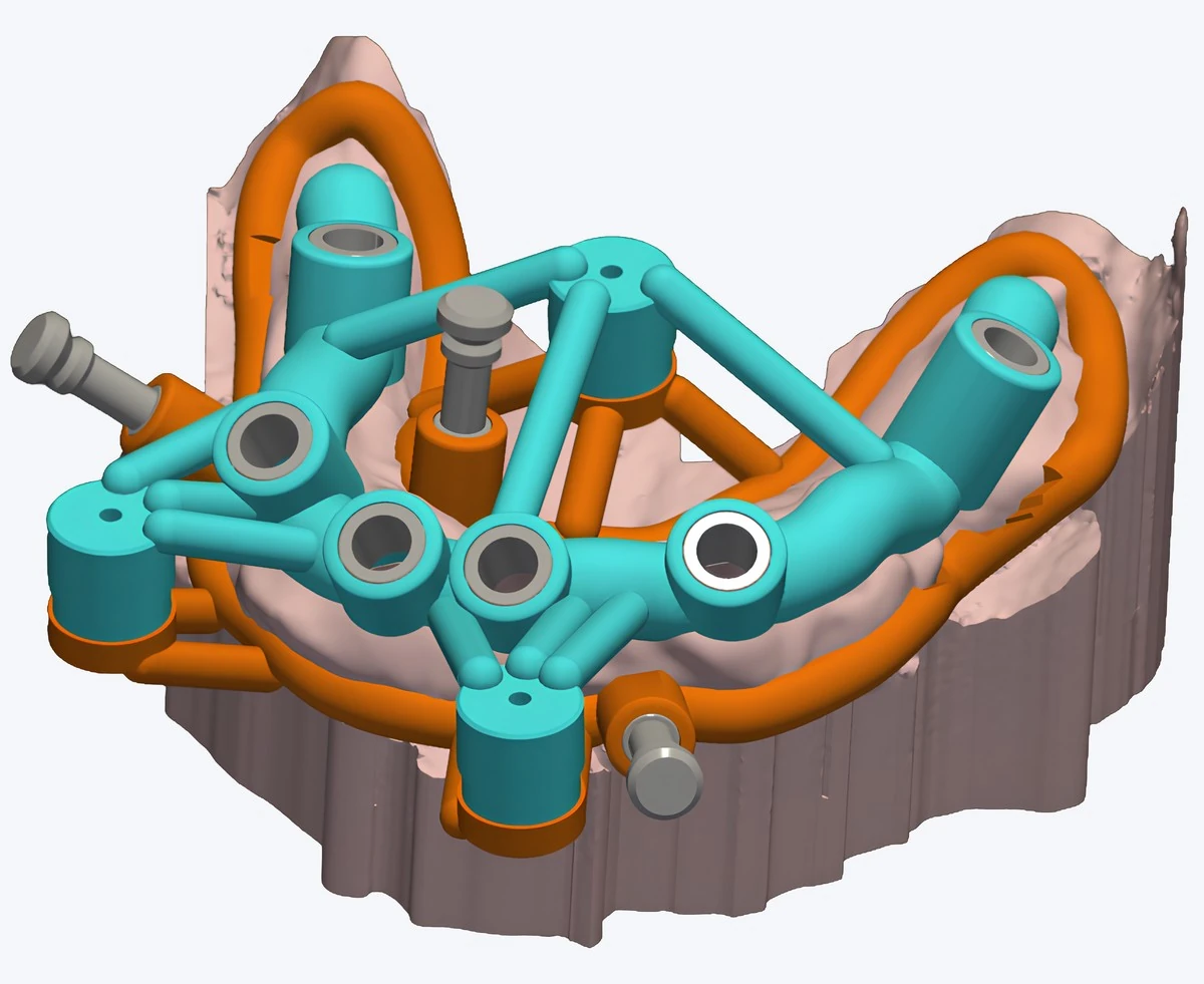
A zygomatic surgical guide seated on the segmented bone, its sleeves holding both trajectories towards the zygomatic body
The hardest case we plan

Fully Guided Zygomatic Implants

When the posterior maxilla has resorbed past anything a fixture can hold, the anchorage moves to the zygomatic body — and the axis that gets there is long, oblique, and passes close to the sinus and the orbit.

  • Both trajectories planned on the CBCT before anything is cut
  • Entry, angle and depth carried by the guide from crest to zygoma
  • Anterior fixtures placed in the bone that is still there, in the same plan
A patient-specific guide seated on the model, its sleeves and fixation pins holding the plan in place
When one guide is not enough

Stackable Guide

A base seated and pinned once, and every guide after it stacks onto that base — reduction, then drilling, then placement. Each stage inherits the position the one before it was planned in, instead of being found again by hand after the ridge has changed shape.

  • One base, pinned to the bone, carrying every later guide
  • Bone reduction and osteotomy share one reference, not two
  • The plan survives the moment the landmarks it was drawn on are cut away
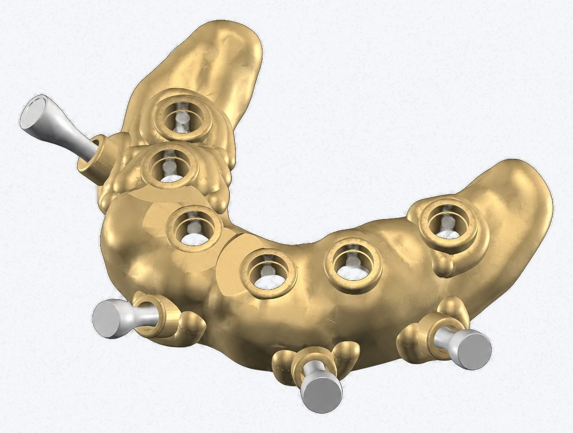
A screw-retained full-arch framework on its multi-unit abutments, seen from above
The case we plan most

Full Arch

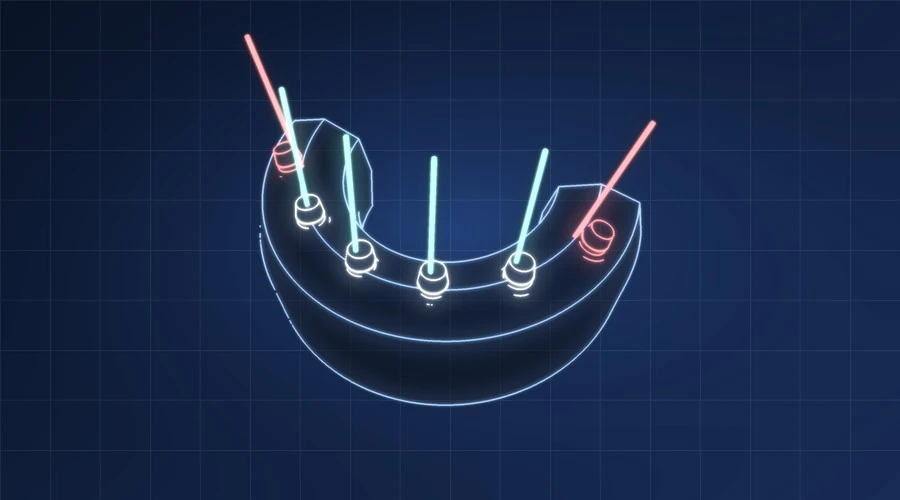
Four to six fixtures carrying one prosthesis. The bar is decided first and the fixtures are placed to reach it — which is what prosthetically driven planning means, and why the posterior pair is tilted rather than dropped into whatever bone is under it.

  • Posterior fixtures tilted away from the sinus and the mental loop
  • Abutment height and angle chosen per fixture, not after the fact
  • One guide carries all of it — entry, angle, depth
Also planned here

Three more case types

A shaped bone graft seated against the maxilla in the position the planned fixtures need
When the ridge has to be rebuilt first

Guided Bone Regeneration

The block is designed from the CBCT to sit where the fixtures are going, and it is placed and fixed in that position instead of being trimmed to fit by hand at the chair.

  • Designed to the planned fixture positions
  • Fixation screws planned clear of the roots and the nerve
  • The implant plan comes back with the graft
See a published case Ask about a regeneration case
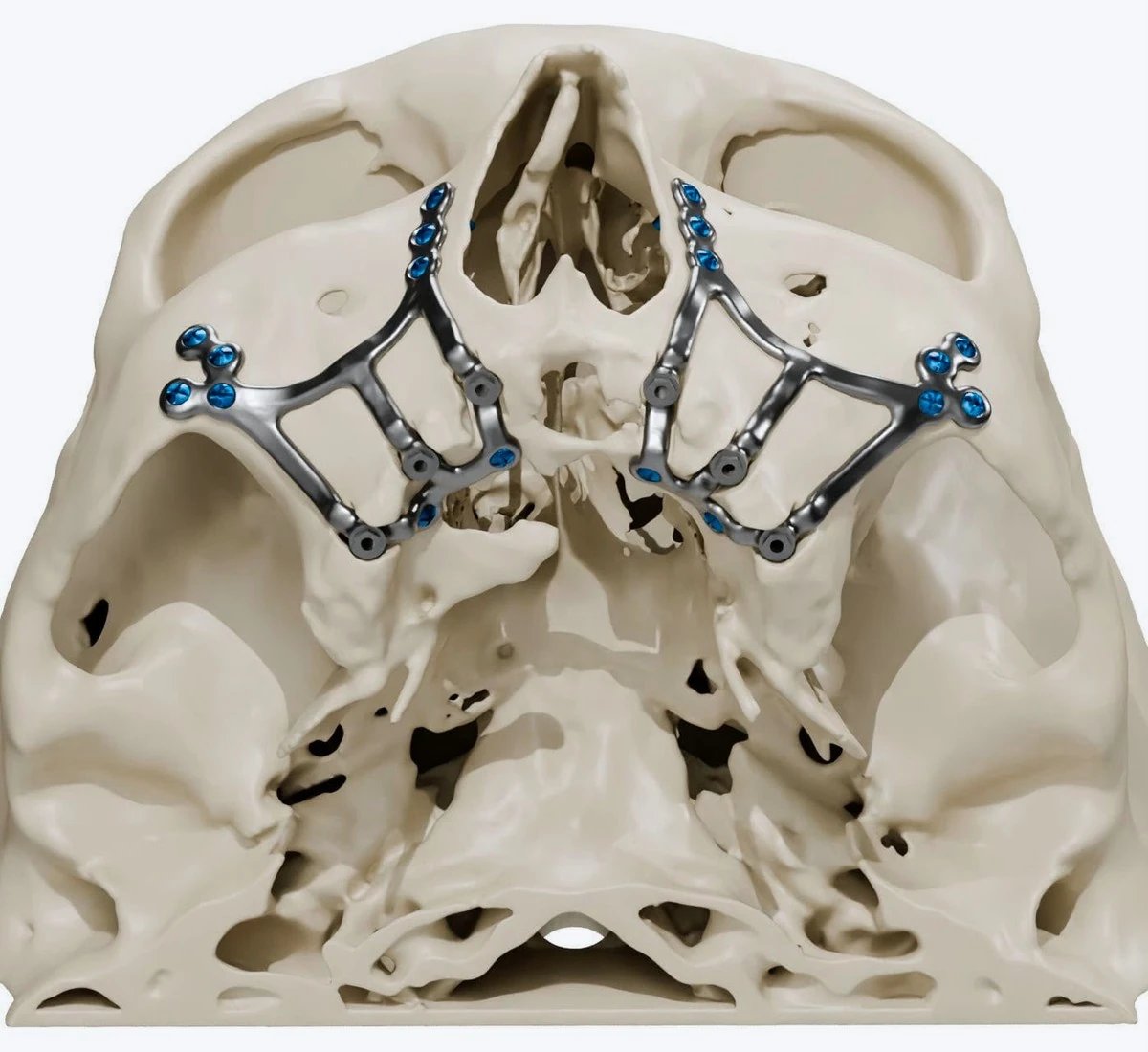
Patient-specific titanium subperiosteal frameworks fixed with screws to both sides of the midface
When there is no ridge left

Subperiosteal Implants

A framework designed for one patient from that patient's CBCT, fixed to the bone that remains and carrying the abutments itself. Nothing goes inside the ridge, because there is no ridge to go into.

  • One framework, one patient
  • Fixation points chosen where bone still exists
  • Abutment positions decided with the prosthesis
See a sample case Ask about a subperiosteal case
A printed reduction guide, its rails setting the crestal level the prosthesis was planned to sit at
Before anything is drilled

Guided Bone Reduction

The crestal level is decided on the plan, where the prosthesis and the bone can be seen together, and a reduction guide cuts to it — so the ridge is brought to the plan rather than the plan re-judged against the ridge.

  • The cut plane comes from the prosthetic plan
  • Pins hold the reference after the landmarks go
  • The drilling guide seats on the same base
See a published case Ask about a reduction case
Who receives your case

Every case that arrives goes to one person, not a queue. He opens your files before the clock starts, tells you the same day if something cannot be planned from, and stays with the case on WhatsApp until the plan is back with you.

Not sure which service your case needs?

Send us the case details on WhatsApp and we will tell you which one it needs.

How it works

From upload to printed guide

Every case moves through the same seven stages. You can see which stage yours is at, at any time, from your dashboard.

The seven stages of a case

  1. 01

    Subscription check

    Your case is uploaded and queued while we check the subscription is active. Nothing is invoiced for the case itself — if the subscription is current, this step passes in minutes.

  2. 02

    Received

    The subscription is confirmed and your files pass a completeness check. The delivery clock starts here — not at upload.

  3. 03

    Missing files

    Only if something is unusable or absent. We tell you exactly what to re-upload. The case is not rejected and it costs you nothing extra — the clock pauses until the files arrive.

  4. 04

    Under review

    A consultant reads the CBCT: bone volume, density, anatomical limits, and the position the prosthetics require. This is a clinician's judgement, not an automated output.

  5. 05

    In design

    For guide orders, a designer builds the guide against the approved plan — sleeve positions, drilling sequence, support and retention.
  6. 06

    Ready

    The report and the guide file are available in your dashboard. Review them and either approve, or request your one free revision.
  7. 07

    Approved / Closed

    You download the files, and 24 hours later everything to do with the case is deleted from the site — your imaging included.

Before you start

What we need from you

  • A CBCT scan covering the full region of interest
  • An STL intraoral scan — required only for a surgical guide
  • Patient name, age, gender and relevant medical history
  • Number of implants, implant system, surgical kit and guide type
  • Confirmation that the patient consented to the upload
  • Your approval of the plan before we finalise it — we send it to you for confirmation and wait
Accepted formatsZIP (DICOM) · STL · PLY · OBJ
Maximum size1 GB per case
Report delivery3 working days
Guide delivery5 working days
Rush service24 hours
Free revisions1
Files kept on the site24 hours after delivery
Files & privacy

How we handle patient data

You are uploading real patient imaging, and that carries obligations for both of us.

  • Every submission requires you to confirm you obtained the patient's consent.
  • Access inside Future Guide is limited to the consultant and designer assigned to your case.
  • Everything is deleted 24 hours after the case is delivered — the imaging you sent and the files we produced. Download them the same day.

The report is consultative and the guide is a planning aid. Final clinical responsibility rests with the treating dentist.

Guided surgery

From your scan to a guide you can print

A consultant reads your CBCT, plans the implants, and designs the guide. You approve it, then print it on the printer in your own clinic.

No software to buy, and none to learn. We plan in Implant 3D — the Italian planning software we distribute across the region.
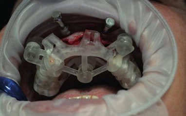
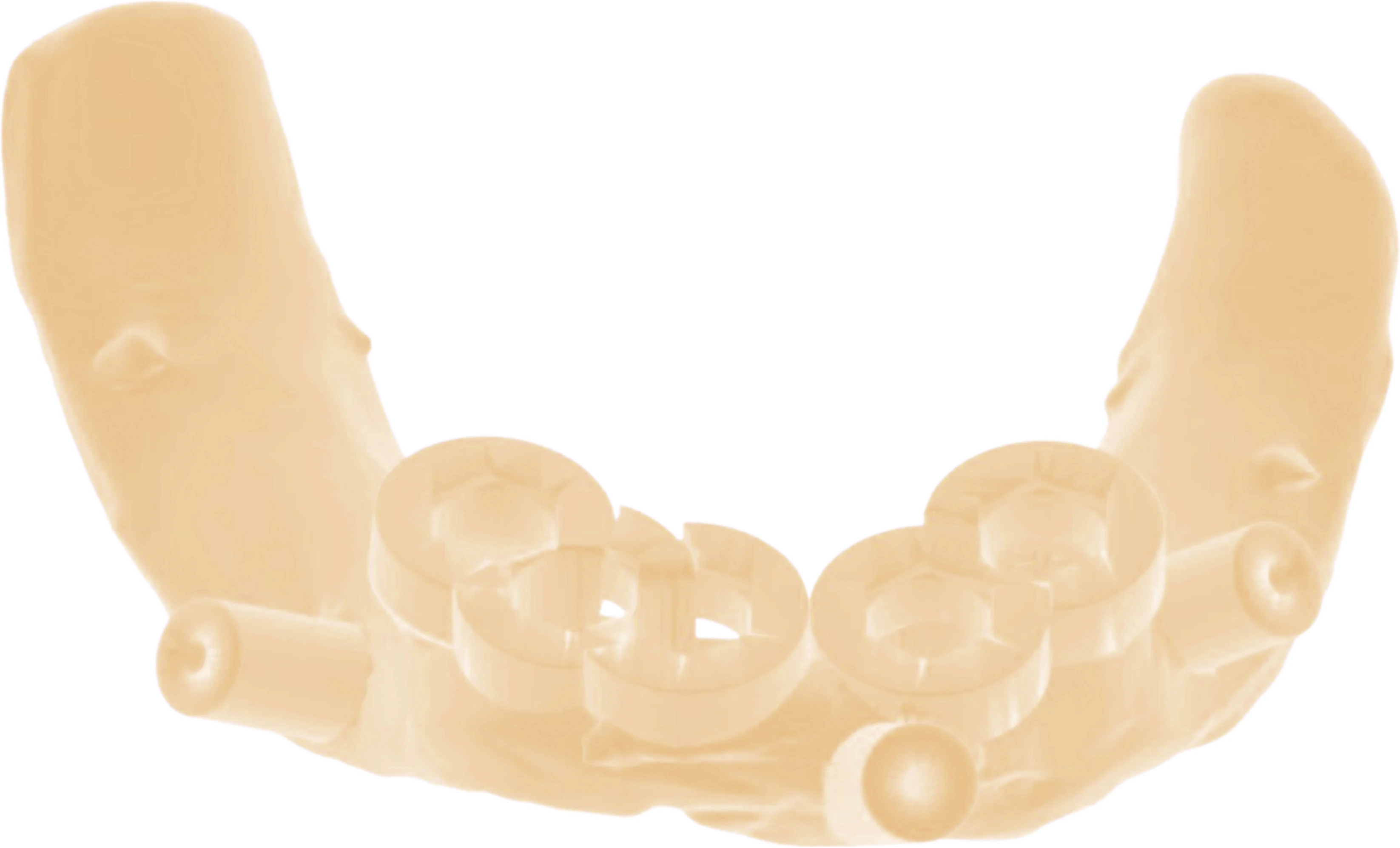
A printed surgical guide seated on an arch model, with the drill sleeves in place Guide produced with Implant 3D · Media Lab S.p.A.
  1. 01

    Submit your case

    CBCT, intraoral scan and the case details — one upload, in your browser.

  2. 02

    Digital analysis

    Bone volume and density read, the nerve canal traced, the anatomical limits marked.

  3. 03

    Implant planning

    Position, angle, diameter and length decided by what the prosthesis needs.

  4. 04

    Guide design

    Sleeves, drilling sequence, support and retention — built for your implant system.

  5. 05

    You approve, then print

    Nothing is finalised before you agree with it. Then the STL is yours.

Case types

The cases we plan

Find yours, and see what the plan has to solve before anything is drilled.

These describe clinical situations, not patients. No case, photograph or outcome on this page is presented as work already completed.

The planning software

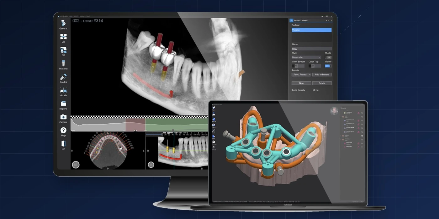
Your case is planned in Implant 3D — the Italian software behind guided surgery

The implant positions, the sleeve heights and the guide itself all come out of one file. Nothing is drawn twice, so nothing can disagree with anything else.

Implant 3D planning software — a CBCT with implants placed, and a stackable guide in Implant 3D Sculpt
Implant 3D and Implant 3D Sculpt are products of Media Lab S.p.A., Italy. Future Guide is their distributor in the Middle East. Software images courtesy of Media Lab S.p.A.

Ready to send a case?

We confirm receipt the same working day.

Contact

Talk to us

We answer every day from 08:00 to 22:00, and reply to written enquiries within 24 hours.

Send a message

Tell us what you need

Choose a subject so your message reaches the right person straight away.

Commercial Registration1508999
Health Licence1508999

What happens after you write

  1. We read it the same day, between 08:00 and 22:00, and reply within 24 hours at the latest.
  2. If it is a case, we tell you which service it needs — a report, or a report with a surgical guide — before you pay anything.
  3. You send the case through the case portal, not through this form.
  4. Work starts once the subscription is confirmed active. Delivery is 3 working days for a report and 5 for a guide.

Have this ready and we can answer in one message

  • How many implants, and which arch
  • The implant system and surgical kit you use
  • Whether you already have a CBCT, and how old it is
  • Your country — it sets the price and how you pay

Please do not attach patient imaging to an e-mail or a chat message. Imaging belongs in the case portal, where it is handled under the privacy policy.

Do not include patient imaging in this form — upload it through the case portal instead.

Partnership

We are looking for distributors

We hold exclusive Middle East distribution for IML Swiss Dental Implants and RESISTA, and we are appointing country partners now.

The opportunity

Two European systems, one region

Implant systems from Switzerland and Italy, paired with a guided-surgery service that gives dentists a reason to choose them over what they already use.

What we provide

  • Exclusive territory rights for your market
  • Product training for you and your sales team
  • Access to the guided-surgery service for your clients
  • Marketing material in Arabic and English
  • Clinical support from our consultant team
Requirements

Who we are looking for

  • A registered company in the dental or medical supply sector
  • An existing sales relationship with dentists in your market
  • Capacity to hold stock and provide local after-sales support
  • Commitment to a minimum annual volume, agreed per territory

Applications are reviewed individually. We appoint one partner per territory.

Questions before you apply?

Speak to us directly — we will tell you honestly whether your territory is still open.
Case submission

Submit a case

Not saved — finish in one sitting
  1. 1Service
  2. 2Case data
  3. 3Files
  4. 4Review
  5. 5Subscription

Which service do you need?

Both start with a CBCT. Choose a guide if position, depth and angulation must be reproduced exactly.

No consultation to book There is no call and no appointment before you start. The consultant’s opinion is part of the case itself — send the case and it comes back with the plan.

Monthly
One subscription, not a bill per case Any number of cases in the month, any scope, any additional planning. The only extra is a rush case.
How the subscription works
After you send

From upload to printed guide

01

Upload

Send your CBCT — and an intraoral scan if you need a guide — with your case notes. ZIP (DICOM) · STL · PLY · OBJ, up to 1 GB. Scan requirements

02

Consultant review

A clinician reviews the anatomy, available bone and the planned implant positions.

03

Design & approval

We design the guide and send it for your approval. One free revision is included.

04

Download & print

Download the print-ready file and produce the guide at your lab or printer. Files are deleted 24 hours after delivery — download them the same day.

Not sure what “a report and a guide” actually means?

Follow one case from the scan to the printed guide — the measurements, the plan and the four-day clock.

See a sample case

Doctor dashboard

Preview only — not a secure area. Sample data, no real login — anyone who opens this file can view it. Do not place patient data, CBCT files or private pricing behind it until the backend is built.

My cases

Sorted by delivery date — the most urgent first.

Case Patient Service Submitted Due Status

Team console

Preview only — not a secure area. Sample data, no real login — anyone who opens this file can view it. Do not place patient data, CBCT files or private pricing behind it until the backend is built.

Case queue

Sorted by SLA risk. Breached cases first, then those closest to their deadline.

Case Doctor Service SLA Assigned to Stage
Subscription

One monthly subscription. No invoice per case.

You subscribe by the month and send cases. Nothing is charged case by case — the only extra is a rush case, and that is a separate fee added to your subscription.

The subscription figure and the rush fee are being finalised — ask us and we will tell you both before you commit.
Monthly subscription Covers every case you send in the month — report or guide, any scope.
Rush case Delivery in 24 hours instead of the standard time. Charged per case, on top of the subscription.
Everything else No extra charge Scope, number of units, additional planning and one revision are all inside the subscription.
At a glance
BillingMonthly subscription
Charged per caseNothing
Units in a caseNo limit
Additional planningIncluded
Rush — 24 hoursExtra fee
Free revisions1
Files kept on the site24 hours after delivery
VATNot applied
Configure a case

Tell us what the case needs

Set the service, the units and anything extra. There is no total to work out — this builds the case summary you send us.

Case configurator

1 · Service

2 · Units in this case

Number of units
1

3 · Additional planning

All of these are inside the subscription.

4 · Timing

Want the figures before you decide?

Message us and we will send the subscription and the rush fee in writing.

Products & distribution

Exclusive Middle East distributor

We hold regional distribution rights for two European implant systems and the Italian planning software behind guided surgery — and we plan the cases that use them.

Coverage

Where you can order today

Supply is live in these markets. If your country is not listed, ask us — we are appointing partners.

Not listed? We are appointing one partner per territory. We are looking for distributors
Exclusive Middle East distributor
View products
IML Swiss Dental Implants RESISTA Implant 3D

Request a quote

Tell us the system, the components and the quantity. We reply with pricing and availability for your country.

Future Guide Academy

International courses in guided implant surgery

Structured international courses and short modules on planning, handling the guide, and the surgical protocol — taught in Arabic and English by the clinician who reviews your cases, and open to dentists across the region.

Your speakers

Who teaches the course

Dr. Maysara Falih Ibraheem AlMaarof

Dr. Maysara Falih Ibraheem AlMaarof

Owner and CEO, Future Guide

Academician and practitioner specialised in guided implant surgery and full mouth rehabilitation, and an international speaker in the Middle East in digital restorative dentistry. Associate at KHCMS — Kurdistan Higher Council of Medical Specialties.

Giovanni Canino

Giovanni Canino

Founder of IML SA

Founder of IML SA, the Swiss manufacturer behind the IML implant system Future Guide distributes. His work is in implant design — thread morphology, the conometric connection, and medical-grade titanium with bio-active surface treatment — and in building IML as a bridge between European manufacturing and the Middle East. A CrossFit athlete outside the company, which he credits for the habit of finishing what he starts.

Dr. Alessandro Tardani

Dr. Alessandro Tardani

Implant surgery · 3-D planning

Academician and practitioner specialised in implant surgery, with a particular interest in 3-D planning and guided implants. Postgraduate masters in conservative dentistry and endodontics, in periodontics and implantology, and in implant prosthodontics and perioral aesthetics. International speaker on 3-D planning in implantology and guided implant surgery.

Eng. Massimo Ivani

Eng. Massimo Ivani

Founder of Media Lab

Entrepreneur in computer science and dental medicine. A degree in computer science and more than thirty years in the industry, and the founder of Media Lab — the Italian company behind Implant 3D, the planning software every case on this site is built in.

Course accessLifetime
CertificateAutomatic PDF
Exam requiredNo
LanguageArabic & English
FormatRecorded video
International courses

The first cohort opens soon

No dates are published yet

We are recording the first course now. Register below and we will contact you with the dates, the fee and the syllabus before it opens to everyone.

Built to move

The video host is not locked in

Where the videos live has not been decided. The player is one swappable component — changing the host later is a setting, not a rebuild.

Want the course before anyone else?

Tell us on WhatsApp what you want covered — the first cohort is being shaped now.

About us

A planning company, not a print shop

Future Guide plans guided implant surgery for dentists across Iraq and the Gulf, and distributes the Swiss and Italian implant systems those plans are built on.

Draft — client approval required
Why we exist

Guided surgery should not be a privilege of large centres

Planning software, a trained eye and a printer are three separate investments. Most dentists in our region have the clinical skill but not the digital workflow — so cases that would benefit from a guide are placed freehand instead.

Future Guide removes two of those three. You keep the clinical judgement and the patient relationship; we do the planning and hand you a file your own printer can produce.

Legal nameGeneral Future Guide
Commercial Registration1508999
Health Licence1508999
Head officeBousher, Muscat
Founded2026
Team3 core + consultants

Registration and licence numbers are shown on every page of this site. Verify them before you send patient data anywhere — to us or to anyone else.

Leadership

Who reviews your case

Dr. Maysara Falih Ibraheem AlMaarof
Dr. Maysara Falih Ibraheem AlMaarof Founder, CEO and lead consultant

Academician and practitioner specialized in Guided Implant Surgery and Full Mouth Rehabilitation, and an international speaker in the Middle East in Digital Restorative Dentistry. Associate at KHCMS — Kurdistan Higher Council of Medical Specialties.

AM
Dr. Ahmed Mohammed Supervisor — operations and case intake

Handles case intake, doctor communication and the daily queue. He is the person who answers when you message us between 08:00 and 22:00.

Markets

Where we work

We serve Iraq and the Gulf today. Europe and the United States are planned, not active — we would rather tell you that than imply a reach we do not have yet.

  • Iraq — direct supply and case planning
  • Oman — head office and direct supply
  • United Arab Emirates — direct supply
  • Remaining GCC — partners being appointed
Why Future Guide

Four commitments we can be measured against

Swiss and Italian systems

Exclusive Middle East distributor for IML Swiss Dental Implants, RESISTA and the Implant 3D planning software.

A delivery commitment

3 days for a report, 5 with a guide, 24 hours when the case is urgent.

One free revision

Every case includes one revision at no additional cost.

Consultant-led

Plans are reviewed by a clinician — not generated automatically by software.

Send us a case and judge for yourself

One case tells you more about a planning partner than any page of copy.

Sample case

What you actually receive

One case, followed from the moment it is uploaded to the file the dentist prints. Every number below is internally consistent, so you can judge the level of detail before you send us anything.

Illustrative case. It is not a real patient — no patient data appears anywhere on this site.
01

What the dentist sent

A single posterior mandibular site. The dentist wants maximum implant length without approaching the mandibular canal.

Case numberFG-2026-0091
ServiceReport + surgical guide
SiteLower left first molar (36)
Patient54, male
Medical historyType 2 diabetes, controlled
Implant systemIML Swiss
Guide type requestedTooth-supported
Files uploadedCBCT 268 MB · STL 61 MB
Doctor's note

“Adjacent teeth are intact and I want to keep the guide tooth-supported. My concern is the canal — tell me the longest implant I can place with a real safety margin.”

02

What the consultant measured

The canal is traced first, then the safety zone is set, and only then is the implant chosen. Not the other way round.

  • Crest to superior border of the mandibular canal: 15.1 mm
  • Ridge width 2 mm below the crest: 7.4 mm
  • Mesio-distal space between adjacent roots: 10.2 mm
  • Bone quality: D2 — dense cortical, coarse trabecular
  • Safety zone reserved above the canal: 2 mm
MANDIBULAR CANAL 2.0 safety 15.1 mm 7.4 mm SECTION · SITE 36
Cross-section through the site. The canal and the reserved safety zone are established before any implant is placed on the plan.
2.6 mm 11.5 mm Ø 4.2 3.4° distal PLAN · v2
The implant is fitted into what the anatomy allows — 1 mm subcrestal, 3.4° distal so the emergence meets the opposing arch.
03

The plan we returned

Every figure follows from the measurements above — nothing here is a default setting.

ImplantØ 4.2 × L 11.5 mm
Platform depth1.0 mm subcrestal
Angulation3.4° distal
Apex to canal2.6 mm
Buccal / lingual wall1.6 mm each side
Drilling protocolFully guided, IML kit

The report states the reasoning, not just the numbers — including what we would change if the ridge turns out narrower than the scan suggests.

04

The guide that carries it

The guide is designed around the approved plan and the exact drilling protocol of the system you use — then exported as an STL your own printer can produce.

SupportTooth-supported
SleeveØ 5.0 mm · H 9 mm offset
Inspection windows2
ExportSTL · print-ready
SLEEVE Ø 5.0 OFFSET H 9.0 GUIDE · TOOTH-SUPPORTED · STL
The sleeve fixes entry point, angle and depth. The guide does the remembering, so you do not have to mid-surgery.
The clock

Four days, against five promised

The delivery clock starts when the files are complete and the subscription is active, not at upload. Here is how this case ran.

  1. D0

    Uploaded · 08:40

    CBCT and STL uploaded, case data completed, patient consent confirmed.

  2. D0

    Subscription confirmed · 11:20

    Subscription checked as active and the file set confirmed complete. The delivery clock starts here.

  3. D1

    Consultant review

    Canal traced, ridge measured, bone quality assessed, implant selected.

  4. D2

    Guide design

    Sleeve position, support surfaces and inspection windows built against the approved plan.

  5. D3

    Sent for approval

    The dentist reviewed the plan and asked for the platform to sit 0.5 mm deeper — the one free revision.

  6. D4

    Ready · report and guide delivered

    Revision applied, files released to the dashboard, WhatsApp notification sent.

Delivered

Two files, and the reasoning behind them

Everything below appears in the doctor's dashboard, and is deleted 24 hours after delivery.

Consultant report PDF · measurements, reasoning, and what to do if the ridge differs
Surgical guide STL · print-ready on any 3D printer
Invoice PDF · issued automatically

Your case will not look like this one

Every anatomy is different — that is the whole point of planning it. Send us yours and see what comes back.

Practical guide

How to take a CBCT we can plan from

Most cases that stall do so for the same handful of reasons — and every one of them is decided in the two minutes before the scan runs. This is what we look for when a case arrives.

General imaging guidance for planning purposes. Always follow your scanner manufacturer's protocol and the radiation rules that apply where you practise.
Instant check

Check your scan before you send it

Drop the exported file in and the page reads its settings — voxel size, field of view, slice count, format — and tells you whether we can plan from it.

The file is read on your device. Nothing is uploaded, and the patient name inside the file is never read.

Drop the scan here or choose a file The DICOM series (zip or folder) and the digital impression — STL, PLY or OBJ
The five decisions

Everything that matters happens before the scan

01

Field of view — include more than the site

A scan cropped tightly to the implant site is the most common reason we ask for a re-upload. We plan against anatomy, and anatomy sits outside the gap.

  • At least one intact tooth on each side of the gap — the guide rests on them
  • Posterior mandible: the mandibular canal along its course, and the mental foramen
  • Posterior maxilla: the sinus floor and the full height above the ridge
  • Full arch: the whole arch plus both condyles' side landmarks where your protocol allows
02

Resolution — fine enough to trace a canal

Voxel size decides whether we can follow the canal wall confidently or have to leave a wider safety margin — which costs you implant length.

  • 0.2 – 0.3 mm voxel is ideal for implant planning
  • Up to 0.4 mm is usually workable for a single straightforward site
  • Finer voxels mean longer scans — and a longer scan is a greater chance of movement
03

Movement — the one that ruins everything

A blurred scan cannot be sharpened later. It has to be retaken, which means a second appointment and a second dose.

  • Chin rest, head support and temple straps actually engaged, not just present
  • Tell the patient the exact duration and ask them not to swallow during it
  • Teeth apart, tongue resting on the floor of the mouth and still
  • Anxious or elderly patients: consider the shortest protocol that still gives a usable voxel
04

Metal — plan around it before you scan

Scatter from crowns, posts and amalgam spreads exactly where you need to see. It can rarely be removed afterwards, but it can often be avoided beforehand.

  • Remove every removable appliance, retainer, jewellery and piercing
  • Keep the arches apart — cotton rolls or a bite block stop opposing restorations overlapping
  • Use your scanner's artefact-reduction mode if it has one
  • Tell us in the case notes which teeth carry metal — it changes how we read the images
05

Export — send the data, not a picture of it

This is the step most often done wrong, and it is invisible until the file reaches us.

  • Export the original DICOM series — not a screenshot, a JPEG, or the viewer’s PDF report
  • DICOM exports as many files: zip the whole folder and upload one .zip
  • Do not send only your viewer's proprietary format — we may not be able to open it
  • Open the export once before uploading and confirm the whole field of view is there
If you want a guide

The intraoral scan matters as much as the CBCT

A surgical guide cannot be designed from a CBCT alone. The CBCT shows us bone; the STL shows us the surface the guide will actually sit on.

  • Same arch as the CBCT, taken at the same clinical stage
  • Capture the adjacent teeth fully — they carry the guide
  • Include the opposing arch and the bite when the prosthetic plan depends on it
  • Export as STL or PLY
  • No intraoral scanner? A desktop-scanned model or impression works — tell us which it is

If the STL and the CBCT were taken weeks apart and the mouth changed in between, the two will not match and the guide will not seat.

Before you upload

The sixty-second check

Free download

Keep it next to the scanner

A printable PDF of this guide and the checklist, in Arabic and English — put it where the scan is taken.

One email with the file. We do not add you to anything.

Not sure your scan is usable?

Check it here in a second without uploading it, or send it on WhatsApp and we will tell you honestly whether we can plan from it.